
If there is one protein we can say we know inside and out, it is the humble lysozyme, which we carry in our tears. This is my first Structure of Christmas for 2016.
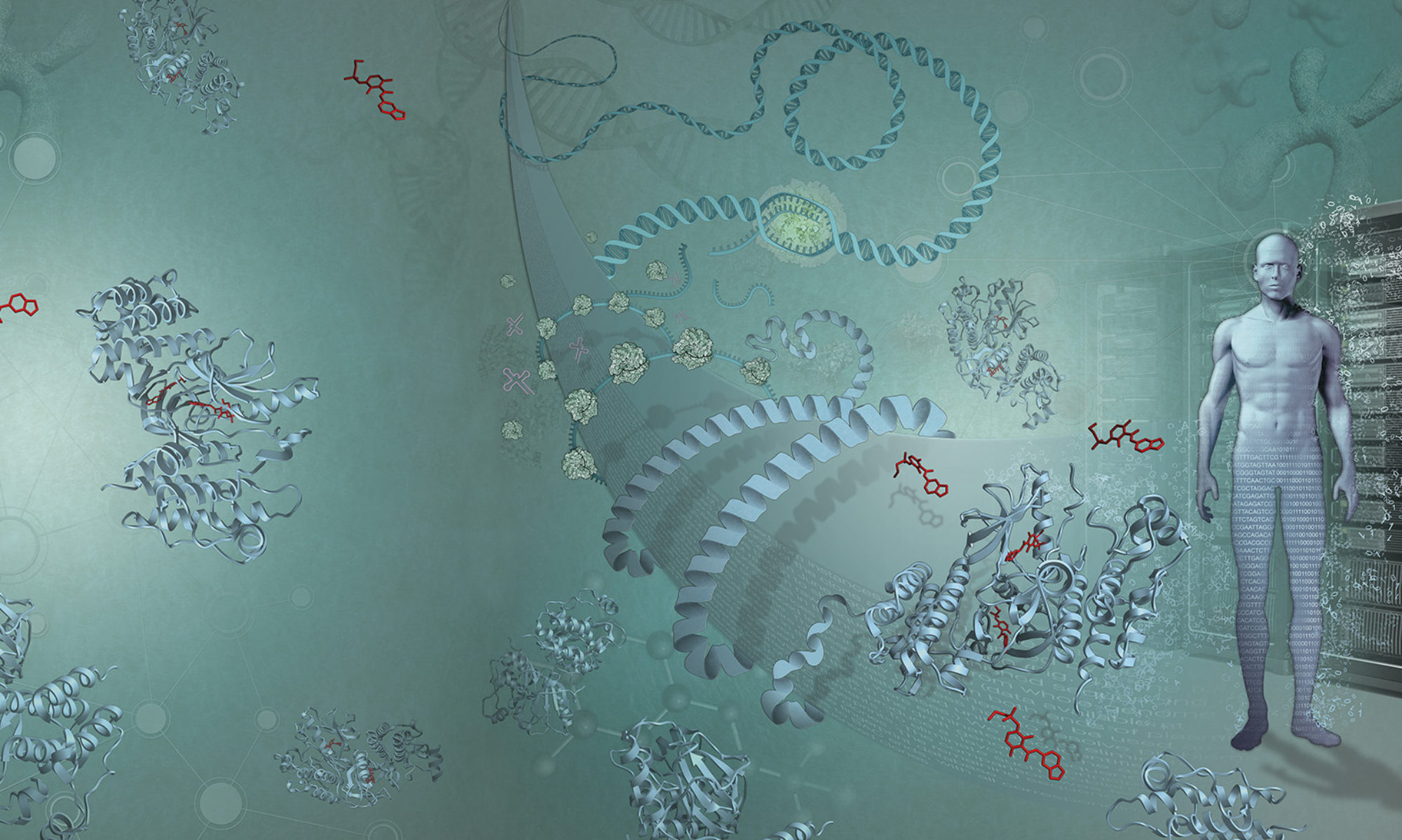
Nowadays, we can see the position of atoms as if we had vision millions of times sharper than human eyes allow. This is the gift of structural biology – the science of determining the 3D structures of molecules and correlating them with function. Structural biology gives us something more powerful than human vision, as the wavelength of ‘visible’ light is too long to resolve the position of atoms.
Structural biologists today have their pick of technologies to determine the structure of a molecule, but the mainstay method is X-ray diffraction. Father-and-son team William Henry and William Lawrence Bragg developed the technique while looking for a way to find the position of atoms in crystals of relatively simple chemicals, and won a Nobel Prize for it (Lawrence was 25 years old at the time).
What’s ‘diffraction’?
Diffraction involves passing photons through a crystal, which scatters them to produce a recognisable pattern. High-energy photons like X-rays provide the best resolution, hence ‘X-ray diffraction’.
A crystal is an ordered collection of a defined set of molecules, in a repeat pattern occurring over such a large volume that you can see it with the naked eye. Table salt crystals provide a familiar example, with sodium and chlorine atoms neatly packaged up. But while neat packages are rare in biology, crystals can be coaxed out of far more complex molecules, like proteins.
In a perfect crystal, every instance of an atom has an identical relationship to its neighbours. Because of this arrangement, photons are scattered in a highly regular way when they are passed through the crystal. This scattering is called ‘diffraction’. The resulting pattern (captured on film in the old days and now detected with a digital sensor) is determined by:
- the relative positions of the atoms;
- the number of electrons they have; and
- how well ordered they are.
Lawrence Bragg was the first person to realise the potential of diffraction and worked out the fundamentals of the process. A new law in physics, Bragg’s Law, was named after him.
Early structures: scaling up the complexity
Structural biology has expanded our understanding of biology to the atomic scale – and unsurprisingly has been recognised with a large number of Noble Prizes. The work leading up to this really got going after the Second World War, when researchers started to take on increasingly complex molecules. Dorothy Hodgkin determined the structures of penicillin and vitamin B12 using X-rays, and was awarded the Nobel Prize for the latter. Max Perutz and John Kendrew were the first to achieve the seemingly impossible: determining the structure of proteins (Perutz tackled haemoglobin, Kendrew myoglobin).
To achieve their goal, Perutz and Kendrew needed bucketloads of pure protein – and to persuade it to form nice crystals. They also had to work out how to ‘phase’ the X-ray diffraction pattern, as phase information is lost in the diffraction experiment. They solved this by soaking heavy metals into their crystals, which results in slightly different diffraction patterns. Calculating those differences allows you to retrieve phase information.
Perutz and Kendrew won a Nobel Prize for this work. Moreover, John Kendrew went on to found the European Molecular Biology Laboratory (EMBL).
The fighter in our tears
The first enzyme structure to be solved via X-ray diffraction was lysozyme, a protein in our tears and mucus, and present in egg whites in large amounts.
Lysozyme is part of our immune system, and its job is to break up bacterial cell walls – so it protects against pathogens very nicely. It is relatively small, but you can purify massive amounts of it (in particular from egg whites) and it is surprisingly happy to crystallise. Because of this, it has become one of the most studied proteins in structural biology.
David Chilton Phillips resolved the structure of lysozyme in 1965, and elucidating its precise mechanism became the subject of intense research by many scientists. Lawrence Bragg was still an active scientist, overseeing the Cavendish Laboratory in Cambridge (where Watson and Crick – using X-ray diffraction images from Rosalind Franklin – deduced the structure of DNA), when he first turned his attention to lysozyme.
Since then, thousands of students have crystallised it, millions of X-ray diffraction patterns have been collected, and we have come to know this collection of 1300 (non-hydrogen) atoms (double that number if you include the hydrogens) perhaps better than any other.
Gazing into inner space
Some protein structures have resolution down to 0.5Å, meaning we can see each atom precisely, even hydrogens, for example seeing the beautiful rings of phenylalanine, tyrosine and tryptophan amino acids atom by atom. We can even discern the oxygen atoms of individual water molecules if they have been “trapped” around the protein.
LYZ, the gene that codes for lysozyme, is just one of 20,000-odd protein-coding genes in the human genome. Each of these genes encodes for the production of thousands of atoms, which are grouped into amino acids. The precise arrangement of these atoms was ‘chosen’ by evolution to form not just a gloopy mess, but a specific three-dimensional shape. Sometimes that shape is deliberately floppy, but for the most part proteins are precisely structured.
Blink, think, fly…
Proteins do the majority of the work in our bodies – moving our muscles, replicating DNA, allowing ions to act on neurons so we can think… And it all comes down to these precise configurations of atoms. One cannot underestimate the importance of uncovering their structures, or of getting every detail of those structures precisely correct.
It is amazing how evolution has been able to arrange and preserve these elegant collections of atoms – not just for us but for millions of organisms, each of which is home to billions of different proteins. They do everything from digesting uranium compounds in the bowels of the earth to setting the myriad angles of feathers in the wings of birds flying above us.
Next up…
In the following blog posts I will take you on a tour of a number of these protein structures. We have a 3D model for each of the next eight structures, made for the Christmas trees at EMBL-EBI. I’ve written these posts with the wonderful support of the PDBe team and Mary Todd Bergman (as ever). It’s been real fun choosing the structures together and writing about each one. These are only nine of the hundreds of thousands of structures that we know of, and millions that occur in Nature. It is a remarkable world.