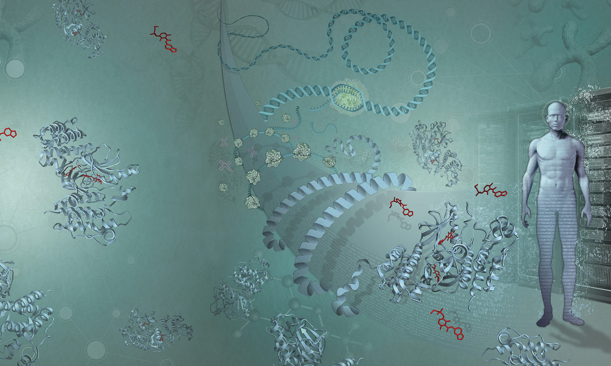
The second structure of Christmas is the membrane protein opsin, which allows us to perceive light.
Proteins that control the information going in and out of our cells are harder to crystallise than run-of-the-mill globular proteins, as they have both water-loving and fat-loving parts and are tricky to mass produce. Opsin, our second structure of Christmas, is one such molecule. It is situated on a special membrane in a specialised cell at the back of our eyes, and senses light.
What keeps us together
The molecules of life are concentrated and organised, in the form of cells. Cells are collections of interesting stuff encased in an incredibly thin membrane: picture tiny soap bubbles filled with a protein drink. We (and indeed all life forms) are walking collections of these over-filled soap bubbles.
That diaphanous layer holding each cell together is the site of intense activity: sensing, interacting and selectively importing or exporting molecules from elsewhere. All of this activity is driven by membrane proteins – few of which are well understood.
Membrane proteins control the information (i.e., signals and molecules) that go in and out of our cells, so it will come as no surprise that most of the known drug targets are membrane proteins. Knowing the exact structures of these proteins is, therefore, intensely important to a lot of scientists.
Border control: a brief tour
Cell membranes are made of two rows of ionic lipids. On the surface, the water-loving part faces outwards; the fat-loving part forms the inside of the membrane. Jostling for space with these lipids are a host of different proteins which span the two rows, with functions specific to sensing and moving things in or out of the cell.
Eukaryotes – all the organisms more complex than bacteria and the leftovers from the primordial soup – have complex cells with organised internal structure. These have additional internal membrane compartments, like soap bubbles within soap bubbles, each of which has its own management regime.
Within each membrane sit a variety of proteins suited to both a watery world (outside the membrane) and a fatty world (inside the membrane). Thankfully, there are many water-loving and fat-loving amino acids to select from amongst the 20 genetically encoded amino acids which make a protein, and a number of elegant molecular gymnastics manoeuvres to fold these proteins straight into the membrane as they are made.
Easier said than done
If you thought working out the structure of a ‘normal’, run-of-the-mill globular protein was hard, that challenge is nothing compared to membrane proteins.
The main problem is that you need lots of protein to make a crystal, but it is extremely difficult to obtain large amounts of one particular membrane protein, like opsin.
To make a lot of a protein you normally make a piece of DNA that encodes the protein, and to push it into a friendly bacterium with a massive “make lots of this” signal at the start of the DNA fragment. But membrane proteins really want to go into membranes, and since membranes are flat, two-dimensional surfaces, there just isn’t room. When you force bacteria to mass-produce a membrane protein, the bacteria often die, presumably from membrane congestion.
If you are lucky, the bacteria will shuffle the foreign protein into special “I don’t know what is going on” section of the cell (called ‘inclusion bodies’). If that happens, the crystallographer can purify the protein – all the while hoping that these elegant proteins have not been turned into some gloopy mess in the process.
Recently, crystallographers have tried using a virus to mass-produce the protein. The virus infects insect cells, and that cell then buds out parts of its membrane continuously. Also as they are insect cells, the protein is more likely to get correctly decorated (many membrane proteins are decorated with sugars). This often gets around the congestion problem, but insect cells are more delicate than their two-billion-year-old bacterial cousins, and need a lot more looking after.
Almost there…
The problems don’t end with membrane protein production. Once you’ve managed to produce enough, you still need to coax them into forming crystals. The combination of water-loving and fat-loving regions demands a huge amount of fine-tuning, for example adding a sprinkle of detergent to appease the fat-loving regions (detergents also have water-loving and fat-loving parts, and there are many to choose from).
The first membrane protein structure was determined 30 years after the first globular, purely water-loving protein structure was resolved. It was slow work, with many intermediate results, but eventually structural biologists developed a reasonably routine (for structural biologists) process.
We now know the structures of hundreds of membrane-bound proteins.
Opsin is so cool
The structure of opsin is typical of a membrane protein: a bundle of long, helical structures criss-crossing the membrane.
You might wonder, “Why would a light sensor be tucked up in a membrane?” At first glance it seems a bit over-engineered. But being situated on the outside of a cell is useful – particularly for single-cell organisms. The other benefit is our light sensing can plug directly into the membrane machinery that controls nearly all signals from the outside world.
In humans, opsin is set in an internal cell membrane. It is arranged like a stack of pancakes, with proteins poised for photons to hit them. When that happens the helices rotate relative to each other, changing the shape of the interior of the protein. Other proteins sense this change, and trigger the cell to send an electric signal to our neurons.
This happens thousand of times per second across thousands of cells. If you are reading this with your eyes, it’s happening in you right now.
A cell within a cell
Dispatching signals and fielding new ones from the outside world represents only a small fraction of what cell membrane proteins do. One of the most fascinating and important functions, I think, is energy capture.
Mitochondria are the power plants of all complex life, and use a super-impervious membrane featuring a web of large, complex proteins to capture energy. They are ancient bacteria that united with another large cell long ago to form the first complex cell: the eukaryote, the root of the family tree that includes us, and all animals. You can still find a vestigial bacteria-like genome inside each mitochondrion.
As a cell within a cell, each mitochondrion has two membranes around it. Mitochondria take in and “burn” carbon-containing molecules, and couple that process with forcing protons (hydrogen atoms that have lost their electron) out of their internal space. The protons are then let back into the middle of the mitochrondria, but in doing so they are forced to work for the cell.
This dual process is managed by the specialised proteins that span the inner membrane. They allow protons back in using an awe-inspiring feat of evolution: a sort of mushroom-like rotor, turned by the protons rushing in. As it turns, the rotor squeezes one more phosphate onto a diphosphate molecule, making ATP: the energy currency of the cell.
A similar remarkable dance is performed in chloroplasts: another former stand-alone bacterium and the solar-power generators in plants.
Bring more structures to light
Despite our collective success in crystallising many membrane-bound proteins, life is teeming with many others that have eluded detection or not (yet) yielded to our efforts to determine their structure. Given their importance, we have much work to do and countless proteins to describe, quantify, observe and understand.

