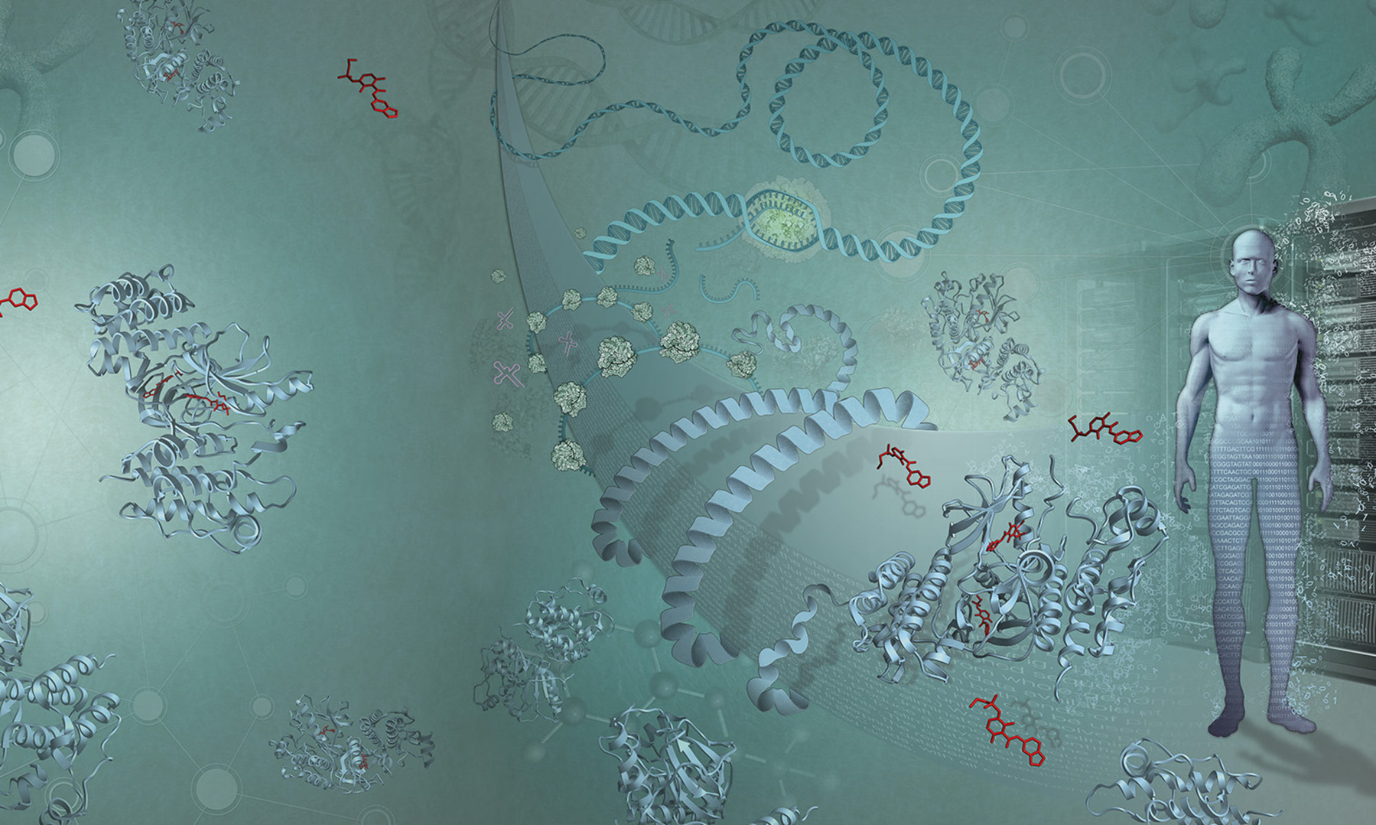
One of the great challenges – and opportunities – over the coming decade is the perfusion of molecular measurement, and accompanying data analysis, into general medicine. This will be nothing new for clinical genetics and other niche disciplines, but as medicine begins to mine the rich data streams from genomics, transcriptomics and metabolomics research, we will start running into some rather tricky integration problems. This is interesting both scientifically and socially, as a huge wave of technology pushes us to create clinical utility out of a confluence of molecular data, high-resolution imaging and data from continuous-sensing devices.
Opinion-makers have been grappling with these issues publicly for a while, and there are programmes in place in many different countries to enable, exploit and empower this change. Futuristic language like “The End of Medicine” and “The Revolution in the Clinic” is bandied about, and governments, charities and companies are all keen to get involved.
I have two different perspectives on thisi ssue. First, as one of the world’s major sources of reference molecular information, EMBL-EBI is a trusted adviser and public data and knowledge provider. Our medical strategy is in place, supported by our advisory boards and ready for implementation (I will be writing a paper on this strategy with Rolf Apweiler). As always, we are prepared to help different sectors and communities deal with ‘big data’ storage, standardisation, integration and knowledge management.
On a more personal level, my research collaborations with clinician scientists have opened my eyes to the challenges and opportunities of practical medicine – some of which I mentioned in my blog post on human as model organism.
I also think we should go back to looking at how different technologies have enriched – but not fundamentally changed – medicine, and at how medicine has adopted new technologies over the years. For me, there is no better example than X-rays (I am indebted to the excellent essay and references from “X-rays as Evidence in German Orthopedic Surgery, 1895–1900”, by Andrew Warwick, Isis, 2005, 96:1–24 )
Technology, medicine and consumers
 |
| Picture of Anna Bertha Röntgen’s hand |
The fact that X-rays could reveal internal aspects of the human body were discovered serendipitously in 1895 by Wilhelm Conrad Röntgen at the University of Würzburg, when his wife accidentally put her hand between radium (a strong X-ray emitter) and photographic film, during Röntgen’s systematic analysis of these new electromagnetic rays. The iconic image of her bones and wedding ring shows at a glance that he had discovered a new way to image living tissue, and in that gestalt moment it seems Röntgen himself understood that this would be useful in medicine.
But it took more than 20 years for X-rays to be used widely in medicine, for a number of reasons. For one, the early developers and adopters of X-ray machines were driven not by medical altruism, but by the need to capture the public’s interest and sell kit – notably in the wealthy, technology-obsessed US at the time. Fair grounds in the northeast began offering ‘bone portraiture’ salons: amusing devices with live radium exposed provided either a picture you could take away or even a fluorescent screen for a live “show”. Such portraits were quite a fad in 1900s New York, with many families proudly mounting pictures of their own X-rays in their houses as a talking point. (This does rather remind me of genomics and genotyping being marketed directly to modern self-obsessed consumers)
 |
| Advertisement for a bone portraiture studio |
There was both enthusiasm and scepticism in clinical circles – mainly the latter. X-rays made bones visible to the naked eye, but didn’t do much else in terms of treatment. The resolution wasn’t good enough to pick up hairline or in-place fractures, and an obviously broken bone didn’t require X rays to diagnose. One German doctor in the 1890s, disillusioned by the complexity in even getting an X-ray machine to work, declared that widespread use of this new technology was “an idle fantasy”. As the poisoning effect of radiation became clear, notably on fairground ‘bone portraitists’, many people in the mainstream medical establishment hardened their view that this technology was mere quackery, useless for clinical practice.
Shooting from the hip
But the ability to see inside the body remained tantalising to many clinicians and scientists, who continued to work on the technology. One group of clinical innovators saw the possibility to improve gallstone treatment. Gallstones are painful, dangerous and difficult to remove, but with the advent of general anaesthetic surgery was becoming a practical option. What was missing was a way to diagnose the presence of gallstones in patients without having to carry out surgery. However, for those keen to make use of X rays, there was a catch. Gallstones, despite being quite solid, were in fact transparent to the ‘hard’ X-rays used at that time – unless they had become calcified, which happened in only 5% of cases. So the clinicians had the right idea: make a better diagnosis to inform a clinical action (surgery) that helps the patient. But a key technical detail made it only occasionally successful. As one might imagine, the anti-X-ray crowd pointed to this failure as indicative of the futility of using the technology at all.
 |
| Modern Xray of childhood hip displacement |
Clinicians were also at the time arguing over whether surgery or manipulation was the best way to treat childhood hip displacement. Manipulating the hip joint into the socket without surgery (under anaesthetic in a medical setting) seemed to work well enough. However, this non surgical approach was traditional, largely carried out by informal medical help, and scorned by the more professionalised medical establishment. As the dispute deepened, the manipulation group (interestingly, led by a surgeon) started taking X-rays before and after treatment to show how the hip joint moved into the correct place following their treatment.
All systems go
Andrew Warwick uses this example to explore how evidence (i.e. X-rays) gains currency in practical medical discourse. Having convinced the medical establishment of the utility of X-rays, more and more practitioners began to buy X-ray machines and engineers began to develop the technology to make and control X-rays in earnest in a clinical rather than physics laboratory setting. Both led to a more widespread adoption for things like orthopaedic interventions and, latterly, the breakthrough use of X-rays to diagnose Tuberculosis.
The use of X-rays was also catalysed by historical events. The Great War called for all manner of medical innovation, and in 1915 Marie Curie and her daughter famously set out to help doctors on the battlefields of France see bullets, shrapnel, and broken bones in their patients in the new Red Cross Radiology Service.
X-rays, radiology and imaging have become essential tools for any medical practice. Every clinician must have at least some working knowledge of the different types of images (and the different risks to the patient). They have practical applications in many disciplines, including neurology, internal medicine and cardiology. The discipline of medical imaging, where rather geeky clinicians work with physicists to push the limits of MRI, X Rays (of all sorts) and echolocation, brings a vast range of technology to bear on improving our ability to look inside the body – and sometimes intervene – in finer and finer detail.
Hindsight is 20-20
Innovative clinicians who believe in the potential of genomics and big-data analysis can learn a lot from the story of X-rays. The enthusiasm of those who grasped the potential of X-rays early on – including visionaries like Röntgen – can serve as a caution, reminding us not to be overconfident about predicting early success. My take-home is that we need to explore many avenues simultaneously – we cannot easily predict where the quickest win will be. Perhaps rare disease diagnosis, or personalised cancer treatment, or infectious biology? But this uncertainty in having to spread our bets to find the first beneficial area should not really deter the long term view that genomics and data analysis will become an every day part of medicine. Fundamentally this is about understanding ourselves at finer and finer detail, and this information will be useful when we are ill. Who can imagine medicine today without medical imaging? Twenty years from now, will medicine before genomics be recognisable?
Direct-to-consumer genomics has enjoyed rapid uptake from early adopters (myself included!) who may be motivated to show off their knowledge, or who may be keenly interested in their ancestry. But the direct-to-consumer market is not the same as integration into healthcare systems; genomics and data analysis is not an “end run” around medical practice, rather another tool for the never-ending quest in trying to ensure our good health.
Just as X-rays and imaging were eventually absorbed and codified into clinical practice, genomics and data science will become so ingrained that we will not remember what it was like before. Medical imaging is a rigorous discipline in its own right but remains firmly rooted in traditional medical structures to ensure it fits seamlessly into practical clinical practice. Because of this, it remains a familiar sight to any patient with a habit of falling out of trees (and their frantic parents).
Similar to medical imaging, genomics and data science will not change the fundamentals of clinical practice; skilled professionals who have seen many similar (but not identical) examples in others can use their experience and knowledge to diagnose and hopefully treat disease. However, it’s quite likely there will be unexpected setbacks and surprising successes in their use, in particular at the start. New medical disciplines (clinical genomics? clinical bioinformatics?) will emerge inside clinical structures, which will provide the
bedrock of routine practice. Every clinician will be expected to have a grasp of the fundamentals of these techniques, and specialists will offer more in-depth knowledge. Society is sure to become more comfortable with this new flavour of information,
and with more self-monitoring (on devices, at home) changing how information is gathered around an individual – those same people who are already more motivated to research on the Internet before the visit to the clinician. But the need for intelligent, skilled individuals who have seen many examples of particular scenario will still be needed to guide, inform and treat, whatever the density of information gathered.
Genomics and data science, once they’ve shown their worth in practical day-to-day practice, will help clinicians make better decisions for their patients. Some diseases will transition from problematic to routine diagnosis and treatment. Some diseases will not be as affected by these technologies. There may be plenty of glitches, dead ends and troubling uncertainties, but if we learn from innovators of the past, using these technologies will quickly become as routine as going to the X-ray department to
examine a hairline fracture.


Hi Ewan,
Thanks for sharing your views on the future role of genomics in medicine.
Some comments on the discovery of X-rays:
Radium is NOT an X-ray emitter. Radium emits alpha particles (He-nuclei), beta-particles (electrons) and gamma rays.
X-rays are generated by shooting high energy electrons on a metal plate. Röntgen discovered this new kind of radiation when he experimented with vacuum tubes and carefully investigated which materials would and would not absorb these rays.
The iconic picture of the hand was made of the hand of the anatomist von Kölliker during the only public lecture Röntgen gave and took an hour to make, so it was hardly an accidental image.
I look forward to your future blogs!
Bram Schierbeek
X-ray crystallographer
OK – let’s get this straight! The radiograph shown above is of Frau Roentgen (https://collections.countway.harvard.edu/onview/items/show/6414) – but is often confused with the later “double ring” radiograph of von Kolliker (https://goo.gl/2mGGWe). The discovery of X-rays and their attenuation by bones was thought to be serendipitous and initially observed using a fluorescent screen. Interestingly, he was colour blind and so he may not have appreciated the effect in quite the same way as his research assistants. The early radiographs required a long exposure with immobilisation of the hand and so were not “accidental” in any sense (http://www.nobelprize.org/nobel_prizes/physics/laureates/1901/rontgen-bio.html).
Genomics and medicine have promoted the development of genetic medicine, which is the development of molecular medicine. First of all, the implementation of genomics, especially the implementation of HGP, the medical home has a new understanding of disease and health.